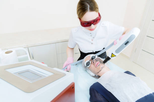
These lasers’ wavelengths (Nd: YAG CO2, erbium, CO2 diode) can affect tissues through ablation, coagulation, and the vaporization process and by stimulating healing processes in cells. Other lasers, with more minor power than surgical lasers, work as “biostimulators.” This chapter explores the most beneficial applications for these lasers, commonly called “therapeutic lasers.” This therapy is often referred to as”low-level laser therapy” (LLLT), but the name is somewhat ambiguous.
The instruments are typically described as “therapeutic lasers” or “cold lasers” in contrast to the “surgical lasers.” Therapeutic Lasers Therapeutic lasers usually occur within the visible red and close-to-visible (IR) portion within the electromagnetic spectrum ranging from 980 to 630 nanometers (nm). The output power typically goes between 50 and 500 milliwatts (mW) using either continuous or pulsed (CW) emitting. The names of therapeutic lasers, as with surgical lasers, are derived from the active medium, such as the gallium-aluminum-arsenide (GaAlAs) laser. The easiest way to categorize therapeutic lasers is to determine their wavelength.
The depth of penetration varies between lasers; those located in the red portion of the spectrum are less absorbent, while IR lasers can penetrate as far up to 3 centimeters according to wavelength and targeted tissue. The laser has an “optical window” at about 820nm, which is the highest optical penetration. Mucosa is very transparent to wavelengths (does not absorb light very well). Skin and bone are excellent, while muscles have the highest absorption of light. The dosage for the targeted tissue has to be calculated according to. Another factor that determines how deep the penetration can be determined is the distance to the tissue, which influences the size of the spot (see Chapter 2). Irradiation outside contact and irradiation that exerts pressure on the tissues provide different doses on the target tissue.
Laser radiation with pressure on the tissue creates a small amount of ischemia in the region, which decreases the hemoglobin levels on the spot (Figures 15-1 and 15-2). Mechanisms The benefit of therapeutic lasers is that they activate natural biological processes. It primarily impacts cells through a reduced oxidation-reduction (redox) process. Cells in the low redox state are acidic; however, after laser exposure, cells become more alkaline and can function optimally.
1 Healthy cells can’t significantly boost their redox levels and cannot react strongly to the radiation energy, while cells in an environment with low redox levels are stimulated.
2 The most significant effect is the growth of the adenosine triphosphate (ATP) that is which is the “fuel” of the cells which is created in mitochondria. Mitochondria.
3 ATP is the end product of the Krebs cycle, in which the photon acceptor enzyme cytochrome c oxidase is inhibited due to Nitric oxide (NO). Laser light dissociates the connection between NO and cytochrome-c oxidase, permitting it to resume ATP production.
4 This fundamental mechanism triggers a chain reaction of cell signaling that leads to better functioning of the body’s functions.
5 Dosage, The most challenging component of LLLT is determining the correct dosage. The dosage for tissue is measured in terms of fluence, also known as energy density which is calculated as joules per square centimeter (J/cm2 ).
Multiplying the power output that the laser produces in milliwatts and the exposure time in seconds is equivalent to the energy. For example, 50 mW x 40 seconds equals 2 000 millijoules (mJ) which is 2.0 J. Once we know the quantity (2 J) and the amount (2 J), we must determine the size of the area that is being irradiated. If we’re illuminating an area of 2 cm2, it is calculated as 2J over a surface of 2 cm2, 2/2 = a flux, energy density, or a superficial dose to give 1J/cm2. Suppose the irradiated area is less than 0.5 cm2. A reverse relationship exists between the spot size (size of the irradiated area) and the fluence. Diminishing the size of the irradiated region boosts the intensity 2. J multiplied by 0.5 cm2 equals 4. The dose then becomes 4 J/cm2 because the energy was distributed in a smaller space, and the intensity was increased locally. Because the quantity depends heavily on the spot’s size, a thin light probe will produce high amounts of J/cm2.However, it does not necessarily mean that the energy transferred to the tissues is significant, and only it means that the light energy emitted from the part of the thin probe is exceptionally high.


